The ear is one of the sensory organs responsible for detecting acoustic stimuli. It also houses the vestibular system. Together with its neural connections, it forms the anatomical foundation of the vestibulocochlear system.
The ear can be divided anatomically into three main parts:
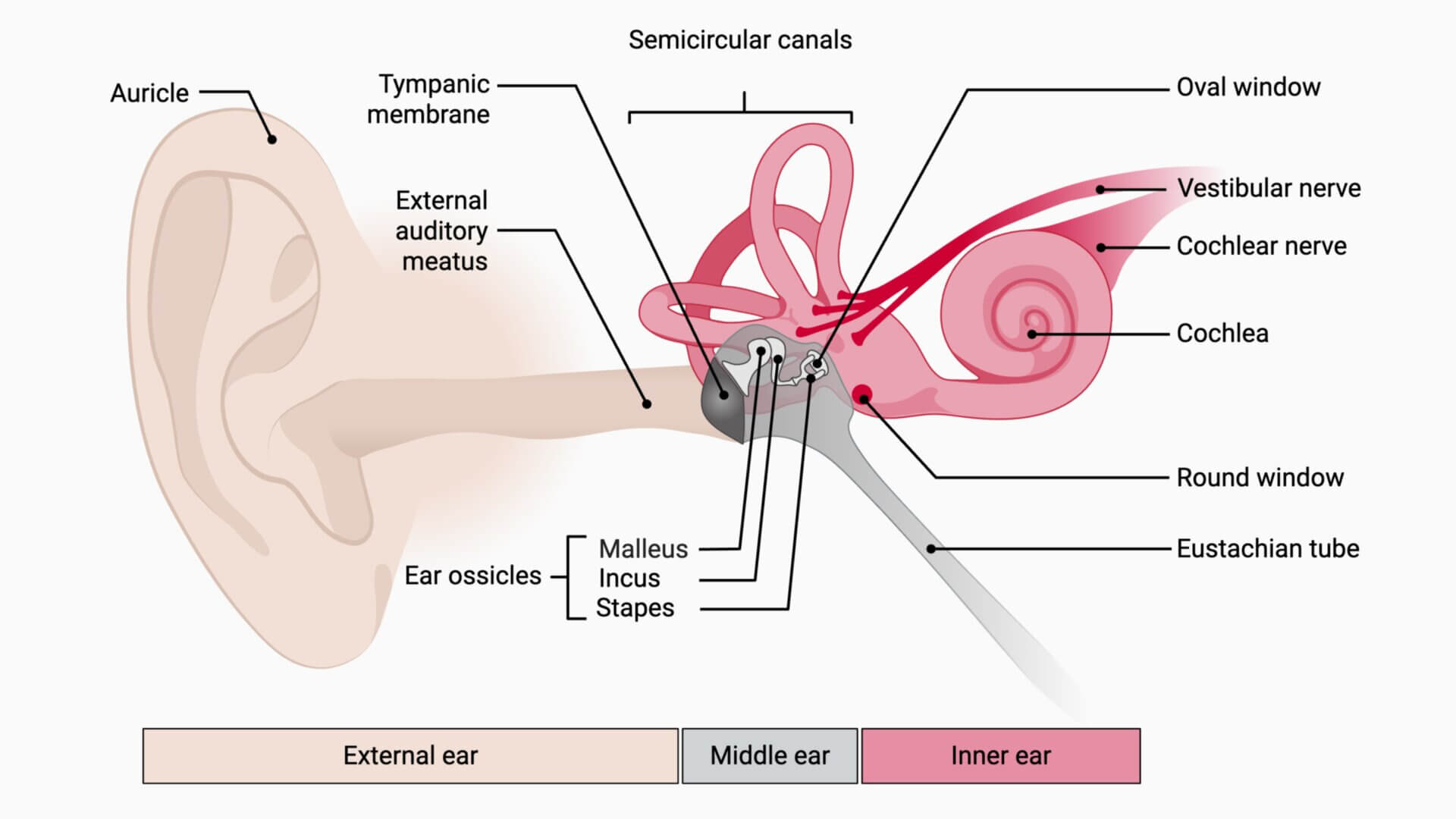
The outer ear consists of the auricle with its distinctive relief elements (helix, anthelix etc.), the earlobe (Lobulus auriculae) and the external auditory canal. Its main function is to collect sound and help determine its direction. The contours of the auricle act as natural acoustic resonators.
The eardrum can be categorized as belonging to the outer ear or the middle ear. It marks the boundary between the two structures. The middle ear consists of a pneumatized cavity in the petrous bone, the so-called tympanic cavity. There are also tympanic sinuses, whereby the air-filled mastoid cells are the most important tympanic sinuses.
The tympanic cavity contains the three auditory ossicles:
In addition, the middle ear muscles are also located in the tympanic cavity:
Other structures in the cavity include ligaments, blood vessels, and nerves. The middle ear connects to the throat via the auditory tube (Eustachian tube), which helps equalize pressure.
The middle ear converts sound from air vibrations to fluid vibrations in the inner ear using the ossicles. This process is called mechanical impedance matching and is necessary because sound travels differently in the air than in the fluid of the inner ear (3000 times higher impedance). Without this adaptation, most sound energy would be reflected instead of transmitted into the cochlea.
The inner ear consists of the labyrinth and the internal auditory canal. The labyrinth is divided into:
There are two main parts of the labyrinth:
The perilymphatic space filled with perilymph is located between the bony and membranous labyrinth.
The cochlear duct contains the sensory receptors for hearing. The vestibular system contains sensory structures that detect head position and motion.
The cochlear duct, saccule, utricle, and semicircular canals are interconnected, forming the endolymphatic system.
Author: Joshua Soeder, DocCheck, created with BioRender.com; adapted from "Middle and Inner Ear Anatomy" licensed under CC BY-NC-SA 3.0