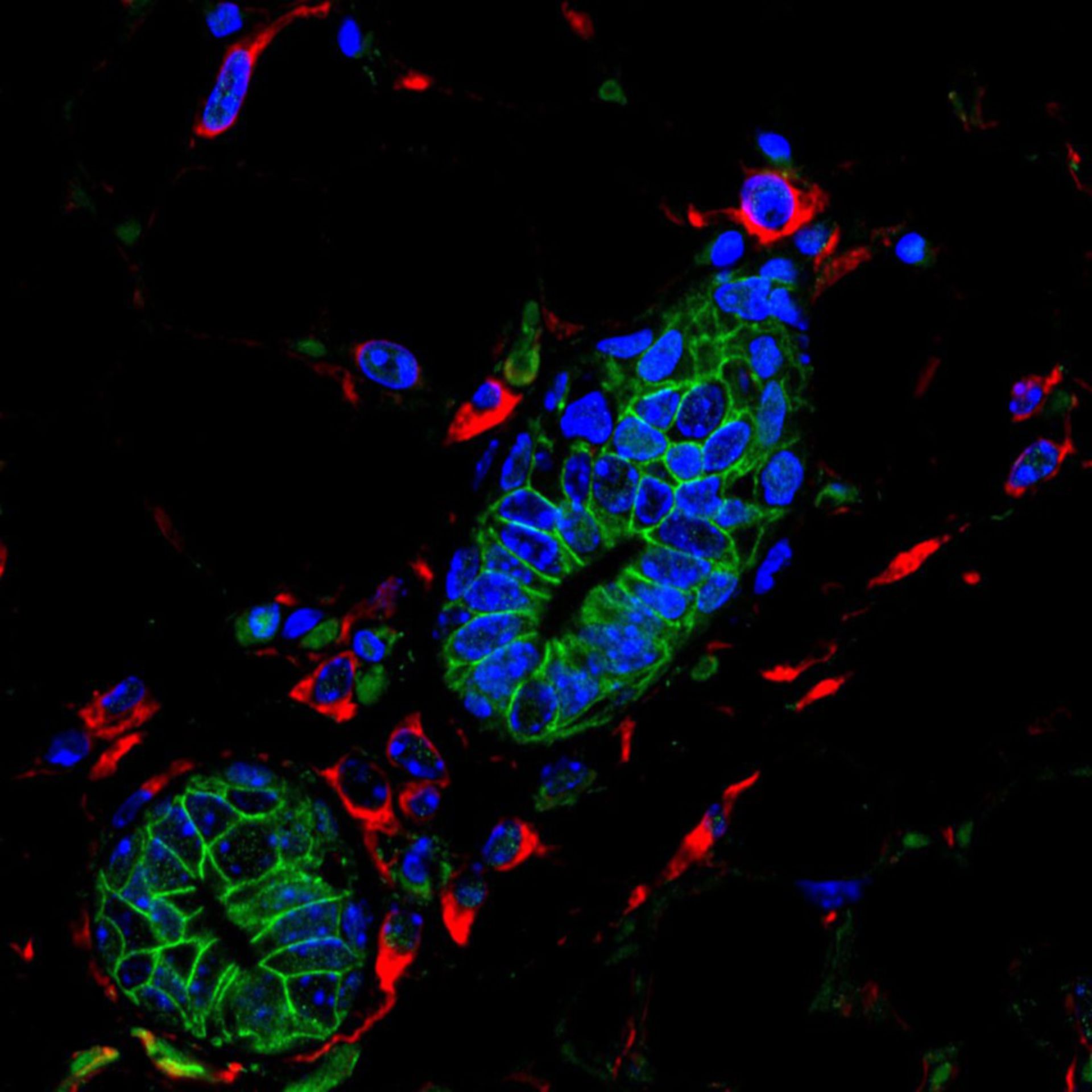
Das Bild zeigt einen Ausschnitt aus einer wachsenden Milchdrüse einer Wildtyp-Maus, die für Vimentin (rot), Beta-Catenin (grün) und DNA (blau) immungefärbt wurde. Dies ist ein Beispiel eines Kontrollbilds für Experimente, die die Funktionen des Elf5-Proteins zeigen. Siehe: Chakrabarti et al. 2012. Elf5 inhibits the epithelial-mesenchymal transition in mammary gland development and breast cancer metastasis by transcriptionally repressing Snail2. Nat Cell Biol 14:1212-1222.
Biologischer Prozess: Laktation
Milchdrüsengewebe aus Wildtyp-P17.5-Mäusen wurde in Formalin fixiert, Wachs eingebettet und Schnitte auf für Vimentin (rot), Beta-Catenin (grün) und DNA (DAPI, blau) gefärbt. Die Bilder wurden mi einem Konfokalmikroskop von Leica aufgenommen. Siehe auch: Chakrabarti et al. 2012. Elf5 inhibits the epithelial-mesenchymal transition in mammary gland development and breast cancer metastasis by transcriptionally repressing Snail2. Nat Cell Biol 14:1212-1222.
Autor: Yibin Kang
Quelle: The Cell: An Image Library