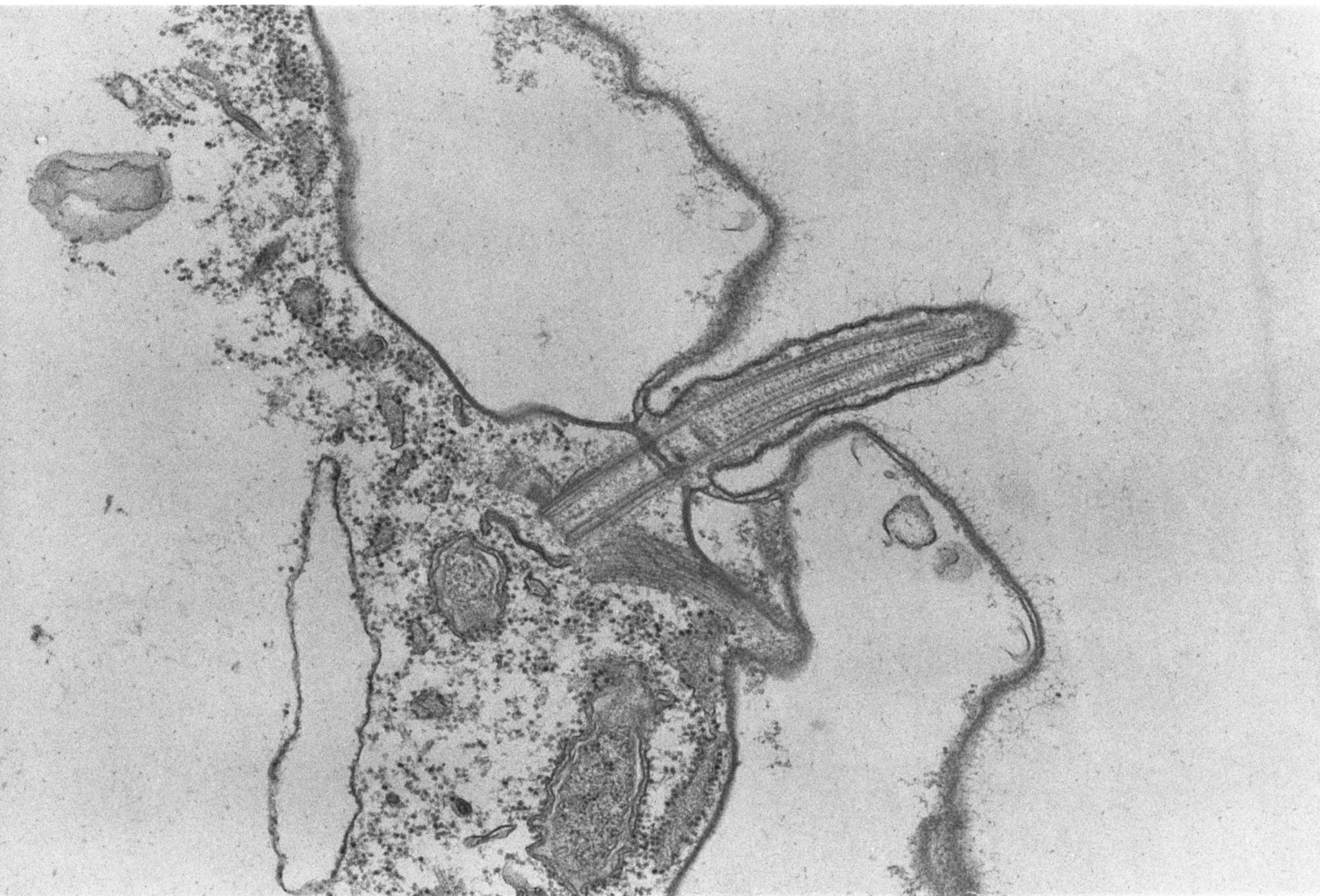
A high resolution image of a longitudinal section of a somatic cilium showing the axoneme, basal body, and attached fibrous and microtubular elements. Standard glutaraldehyde fixation followed by osmium tetroxide, dehydrated in alcohol and embedded in an epoxy resin. Microtome sections prepared at approximately 75nm thickness. TEM taken on 5/24/69 by R. Allen with Philips 300 operating at 60kV. Neg. 18,500X. The raw film was scanned with a Nikon Coolscan 9000ED. This high resolution image is suitable for quantitative analysis Additional information available at (http://www5.pbrc.hawaii.edu/allen/).
Biological Process: Ciliary or flagellar motility, Microtubule cytoskeleton organization, Microfibril organization, Detection of symbiont
Author: Richard Allen
Source: The Cell: An Image Library