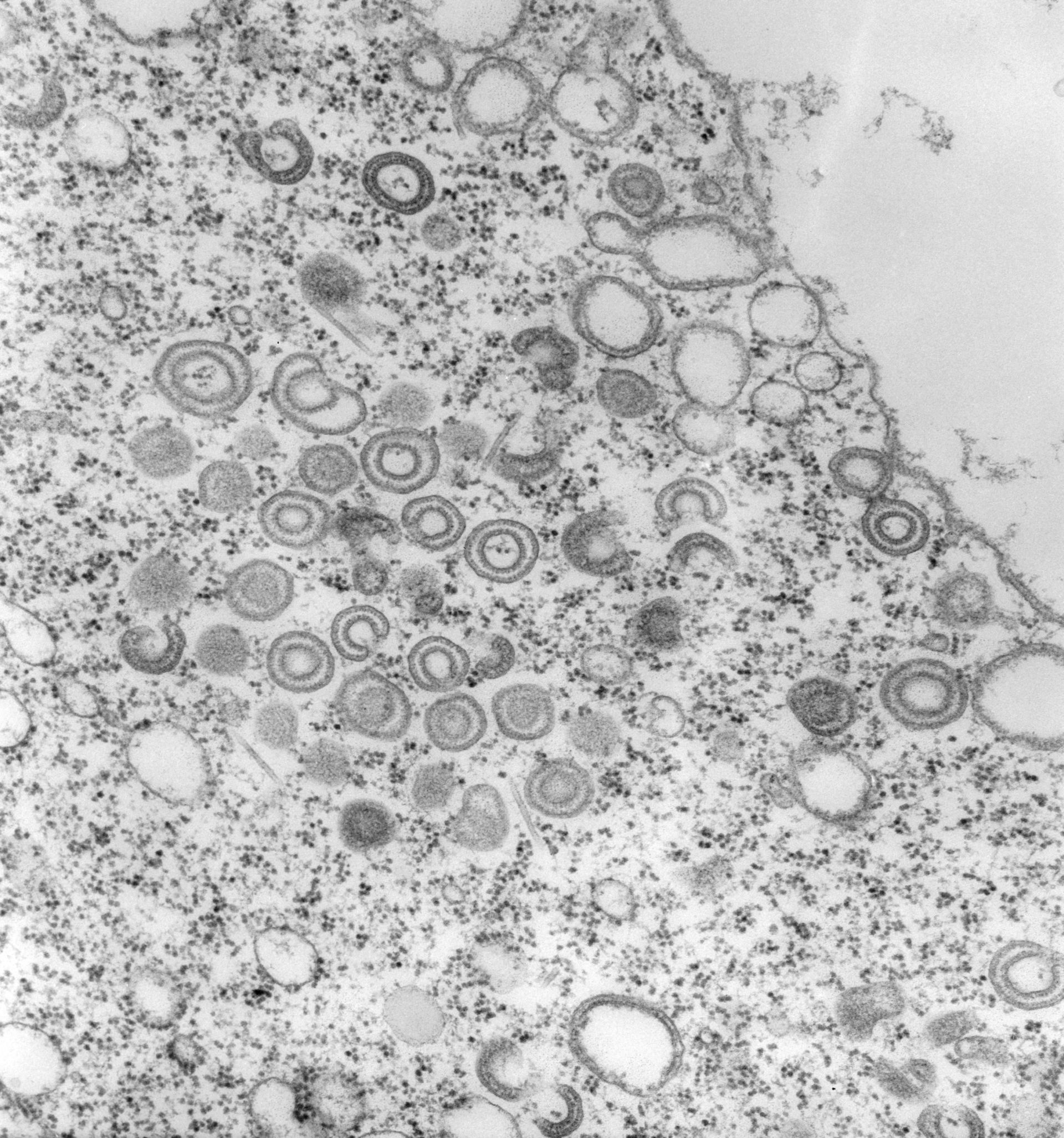
Accumulation of golgi cupulas near a food vacuole. The food vacuole shown has already fused with many vesicles that have already added their membrane as well as their dense inner lining to the vacuole lumen. TEM taken on 2/17/72 by R. Allen with Hitachi HU11A operating at 75kV. Neg. 19,500X. The raw negative was scanned with an Epson Perfection V750 Pro and this high resolution image is best used for quantitative analysis. Additional information available at (http://www5.pbrc.hawaii.edu/allen/).
Biological Process: Post-Golgi vesicle-mediated transport, Golgi to vacuole transport
Standard glutaraldehyde fixation followed by osmium tetroxide, dehydrated in alcohol and embedded in an epoxy resin. Microtome sections prepared at approximately 75nm thickness. Additional information available at (http://www5.pbrc.hawaii.edu/allen/).
Author: Richard Allen (University of Hawaii)
Source: The Cell: An Image Library