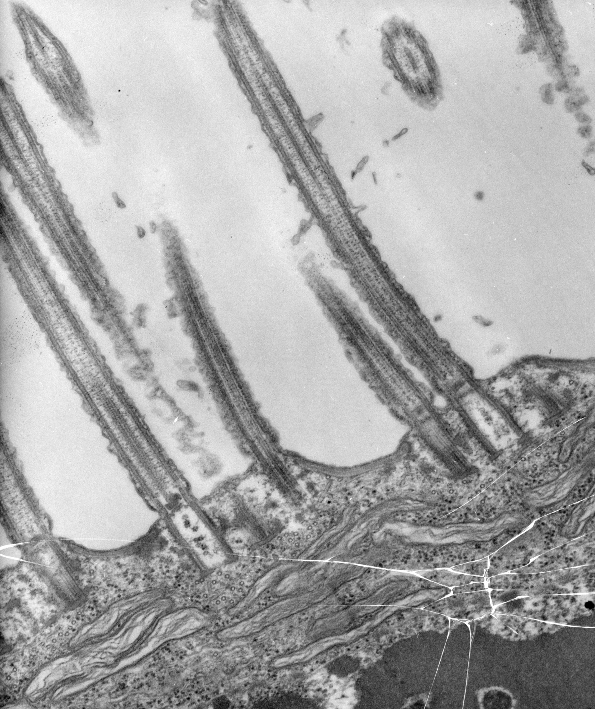
A view of the AZM with basal bodies in longitudinal section and cilia sectioned lengthwise. Bunches of microtubules reside below the basal bodies and lie close to the cytopharyngeal vesicles. TEM taken on 8/3/67 by R. Allen with Philips 200 operating at 60kV. Neg. 19,200X. The raw negative was scanned with an Epson Perfection V750 Pro and this high resolution image is best used for quantitative analysis. Additional information available at (http://www5.pbrc.hawaii.edu/allen/).
Biological Process: Ciliary or flagellar motility, Microtubule basal body organization, Cytoplasm organization
Standard glutaraldehyde fixation followed by osmium tetroxide, dehydrated in alcohol, and embedded in an epoxy resin. Microtome sections prepared at approximately 75nm thickness.
Author: Richard Allen
Source: The Cell: An Image Library