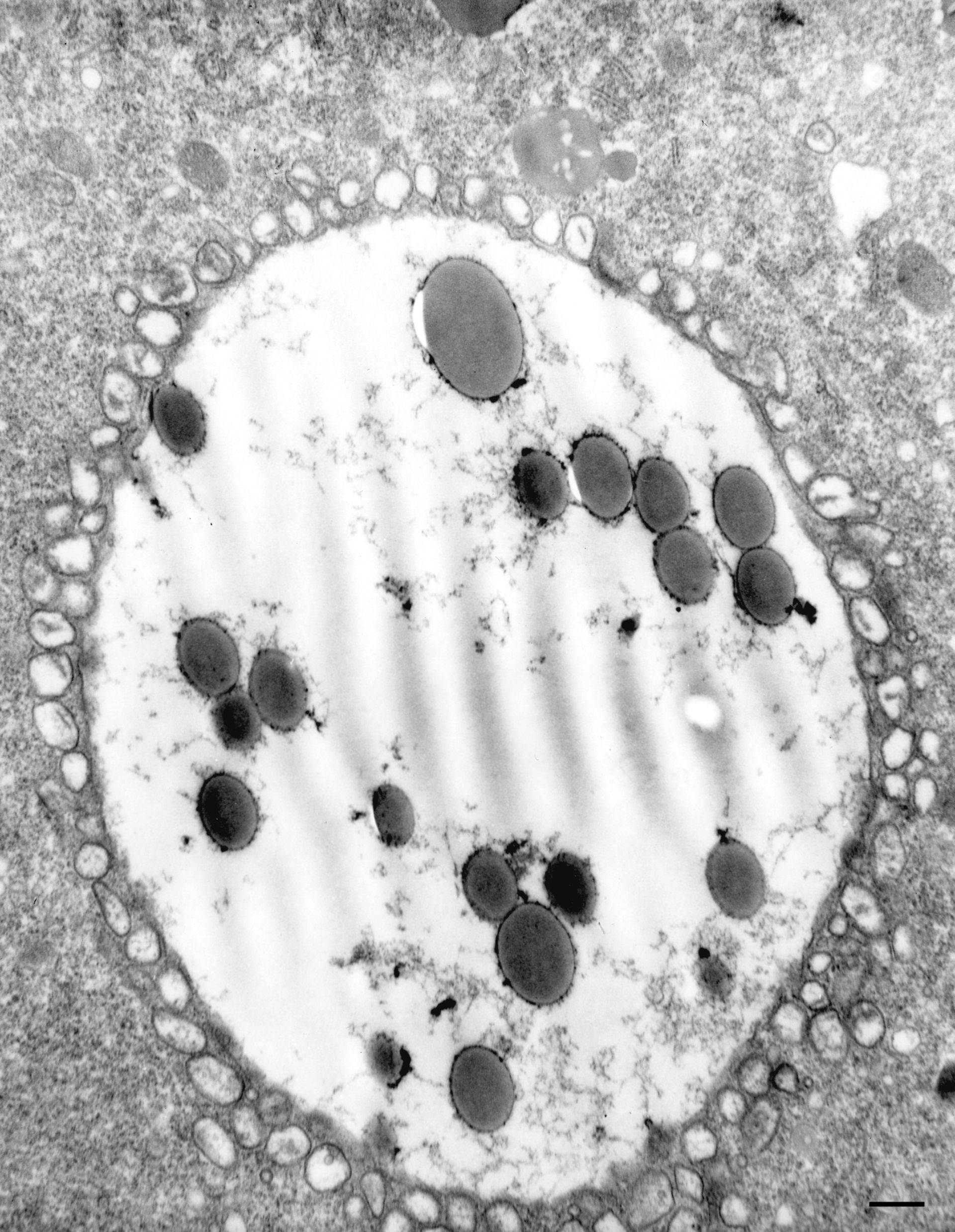
In order to study all stages of DVs in the same cell we used a protocol of sequentially exposing cells to different sizes of latex beads interspersed with washes: for example, 3 minutes in 0.1?m beads, wash 3 min, 3 minutes in 0.3?m beads, wash 3 min, 3 minutes in 0.6?m beads, wash 3 min, 3 minutes in 0.8?m beads, wash 3 min and finally 3 minutes in 1.1?m beads followed by fixing the cells. Thus the 1.1, 0.8, 0.6, 0.3 and 0.1?m bead-containing vacuoles will be 0-3 min, 6-9 min, 12-15 min, 18-21 min and 24-27 minutes old, respectively. The electron micrograph shown here was of a DV-II that was surrounded with docked secondary lysosomes. Secondary lysosomes only recognize and dock at the DV-II membrane. They do not dock with DV-I or DV-III vacuoles. TEM taken on 2/27/81 by R. Allen with Hitachi HU11A operating at 75kV. Neg. 7,000X. Bar = 0.5?m.
Biological Process: Digestive system process
Standard glutaraldehyde fixation followed by osmium tetroxide, dehydrated in alcohol and embedded in an epoxy resin. Microtome sections prepared at approximately 75nm thickness. The negative was printed to paper and the image was scanned to Photoshop. This digitized image is available for qualitative analysis. Additional information available at (http://www5.pbrc.hawaii.edu/allen/).
Author: Richard Allen (University of Hawaii)
Source: The Cell: An Image Library