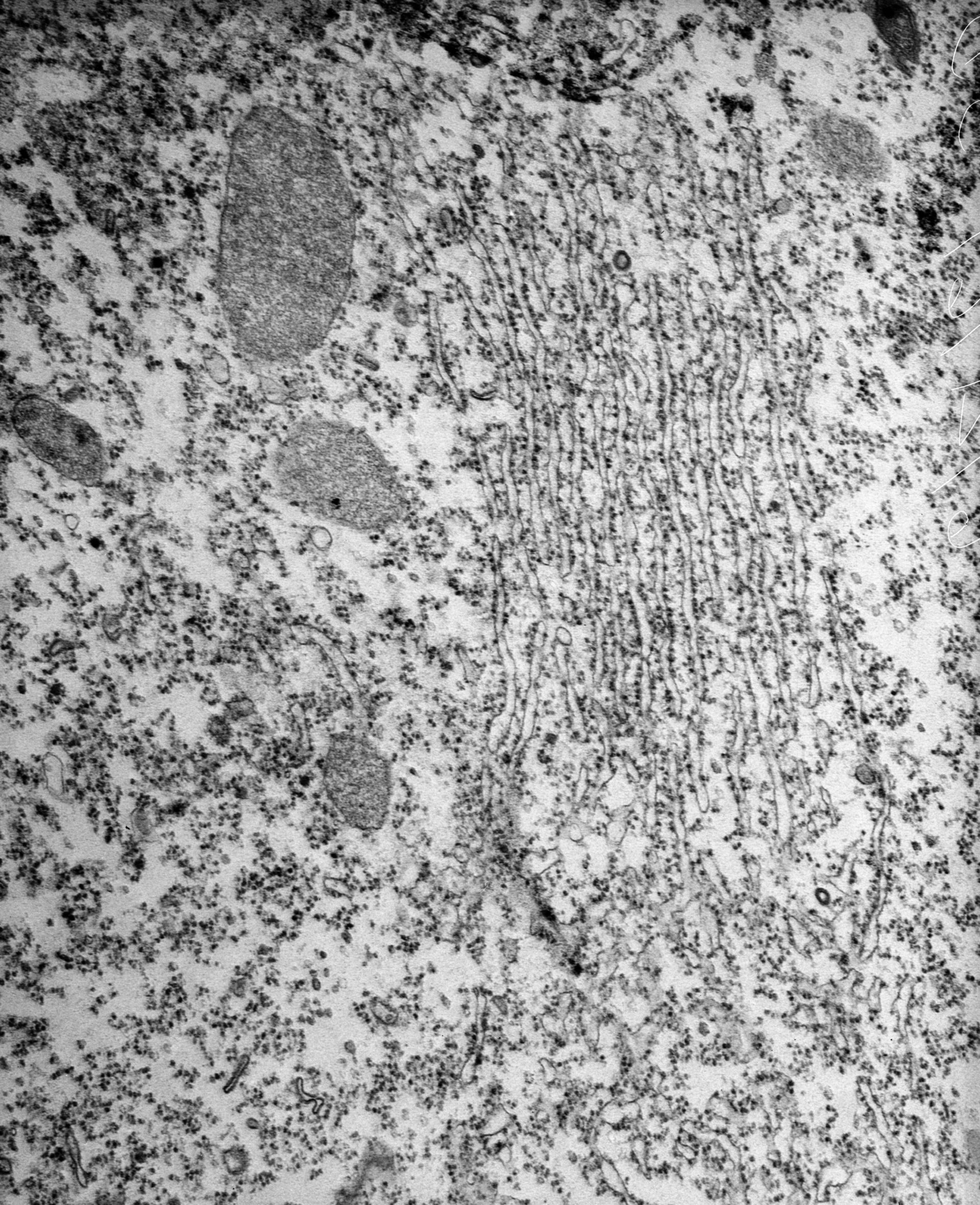
High resolution view of the rough endoplasmic reticulum of Tetrahymena that sometimes forms large stacks or concentric circles from which other organelles are excluded. Peroxisomes lie nearby. EM taken on 6/21/67 by R. Allen with RCA EMU3F operating at 50kV. Neg. 18,800X. The raw film was scanned with an Epson Perfection V750 Pro. This image is best used for quantitative analysis.
Biological Process: Cytoplasm organization
Standard glutaraldehyde fixation followed by osmium tetroxide, dehydrated in alcohol and embedded in an epoxy resin. Microtome sections prepared at approximately 75nm thickness. Additional information available at (http://www5.pbrc.hawaii.edu/allen/).
Author: Richard Allen (University of Hawaii)
Source: The Cell: An Image Library