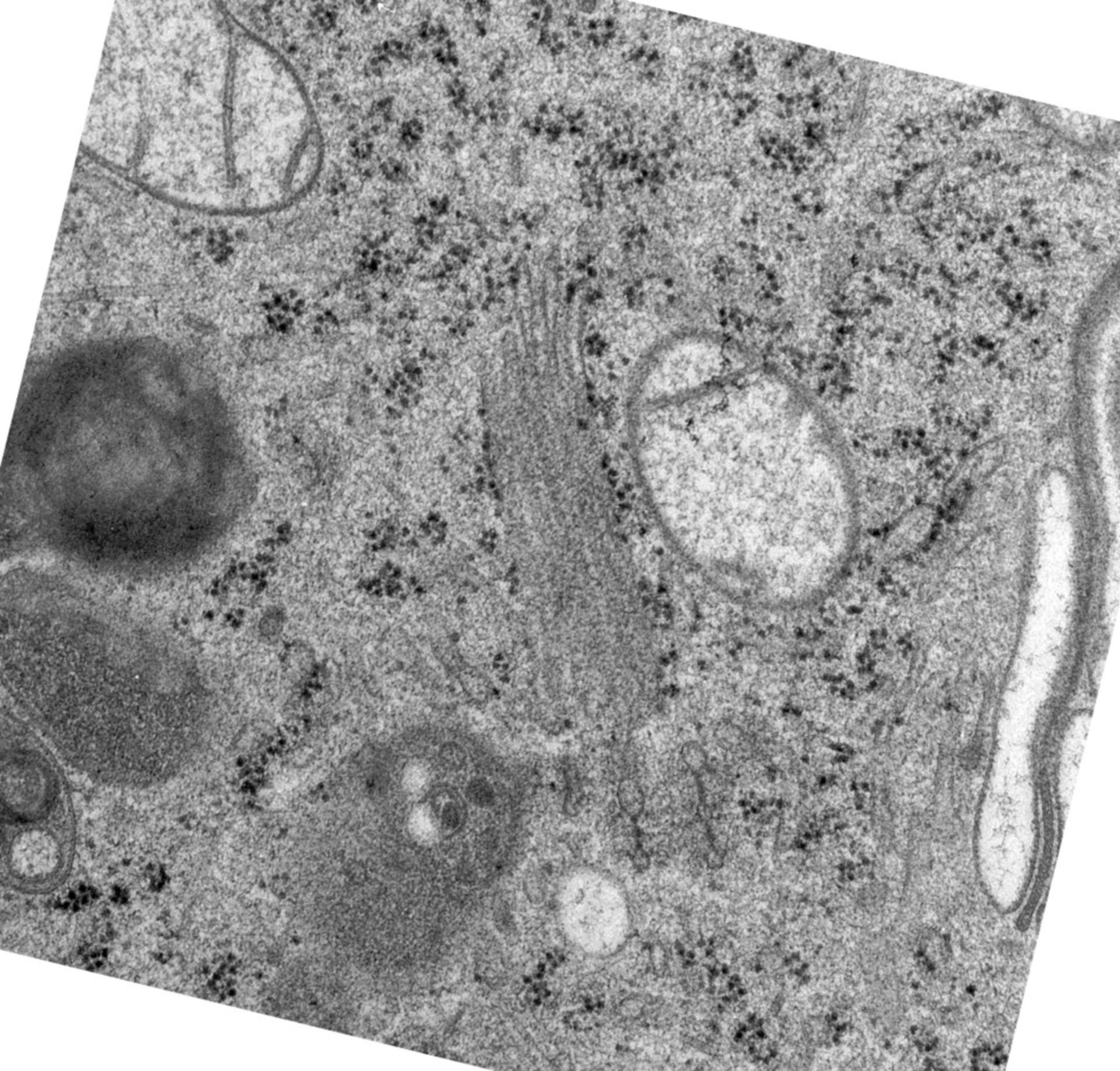
This image is from a tomogram through part of a Golgi ribbon from a normal rat kidney cell, generated using high voltage electron microscopy following incubation at 15?C to block transport out of the Golgi complex. Following temperature block, large bulging domains appear on the three trans-most cisternae. The sample was tilted at angles of 1.5? to obtain the series of images in this set, and used to obtain 3-dimensional reconstructions of the Golgi apparatus and contributed to findings reported in Ladinsky et al. (1999) Golgi structure in three dimensions: Functional insights from the normal rat kidney cell. J. Cell Biol. 144 (6) 1135-1149.
Biological Process: Golgi organization, Golgi vesicle budding
Cells were grown on 100-mesh gold EM grids, and maintained at 15?C for 4 hours before plunge freezing in liquid nitrogen (BalTec HPM-010), followed by freeze-substitution (1% glutaraldehyde, 0.1% tannic acid in acetone, replaced with 4% osmium tetroxide and 0.01% uranyl acetate), then embedded in Epon-Araldite and sectioned at 250nm (UltraCut-UCT, Leica). Sections were transferred to formvar-coated copper-rhodium slot grids (EMS) and stained with 2% aqueous uranyl acetate and Reynold's lead citrate. Digital images were acquired at a magnification of 15,000X with an FEI Tecnai TF20 transmission EM. For additional details refer to: J. Cell Biol. 144 (6) 1135-1149.
Author: L. Andrew Staehelin
Source: The Cell: An Image Library