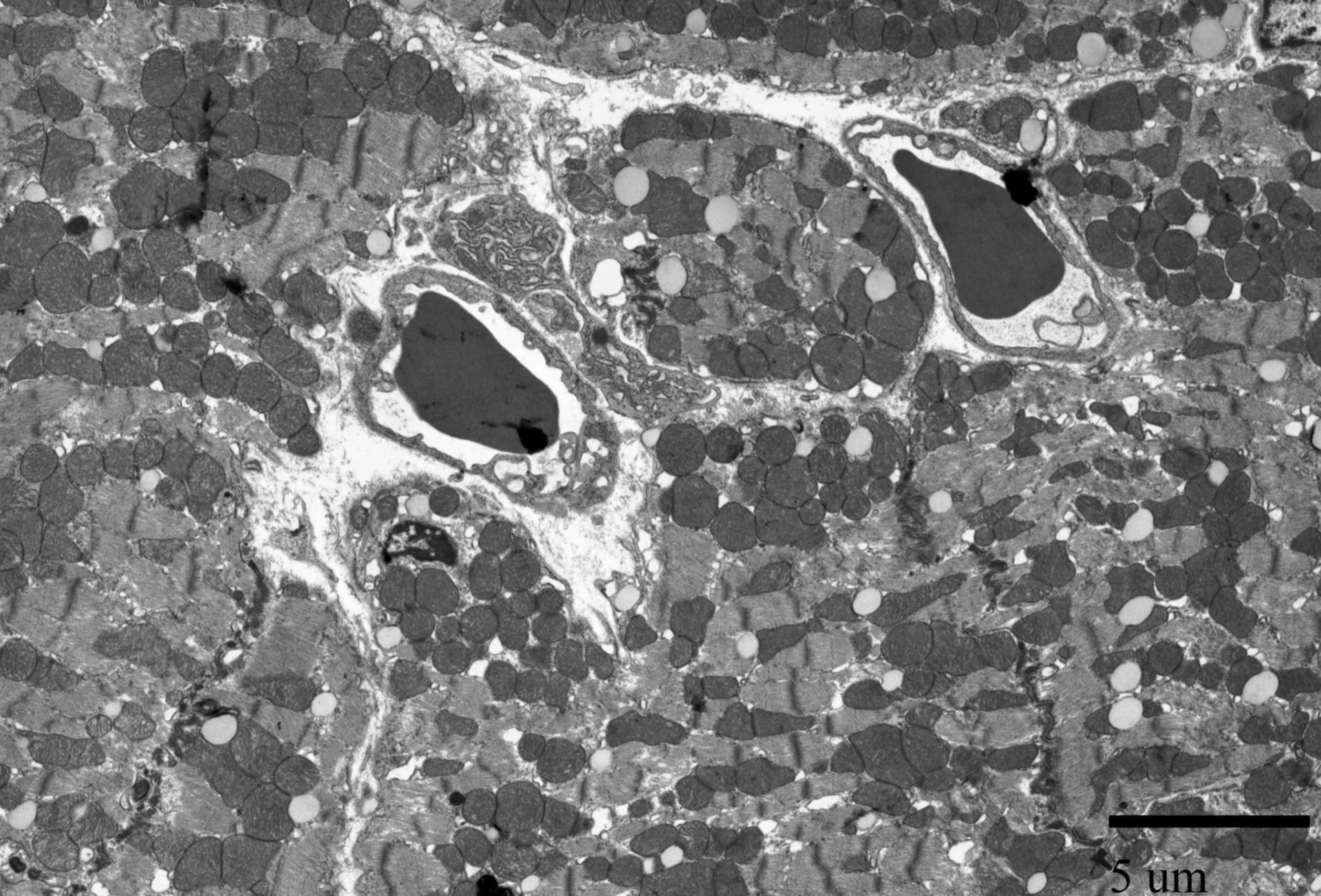
Transmission electron micrograph showing an over view of muscle tissue configuration from a mouse cardiac muscle ventricle. This image shows muscle to muscle connection and the abundance of mitochondria illustrate their importance to heart muscle function.
Biological Process: Cardiac muscle contraction
Primary fixation included: 2.5 % glutaraldehyde, 2% formaldehyde in 0.1 M Na-phosphate buffer, pH 7.4. Post-fixed in 2% OSO4 in 0.1 M Na-phosphate buffer, pH 7.4. Stained en bloc in 1% uranyl acetate. The tissue was then dehydrated in a graded series of ethanol and infiltrated with Spurr’s resin. Thin sections of 70 nm were trimmed using a diamond knife and post-stained in uranyl acetate and lead citrate. This micrograph image was taken using a Phillips CM 100 transmission electron microscope at an accelerating voltage of 80kV.
Author: Marian Rice
Source: The Cell: An Image Library