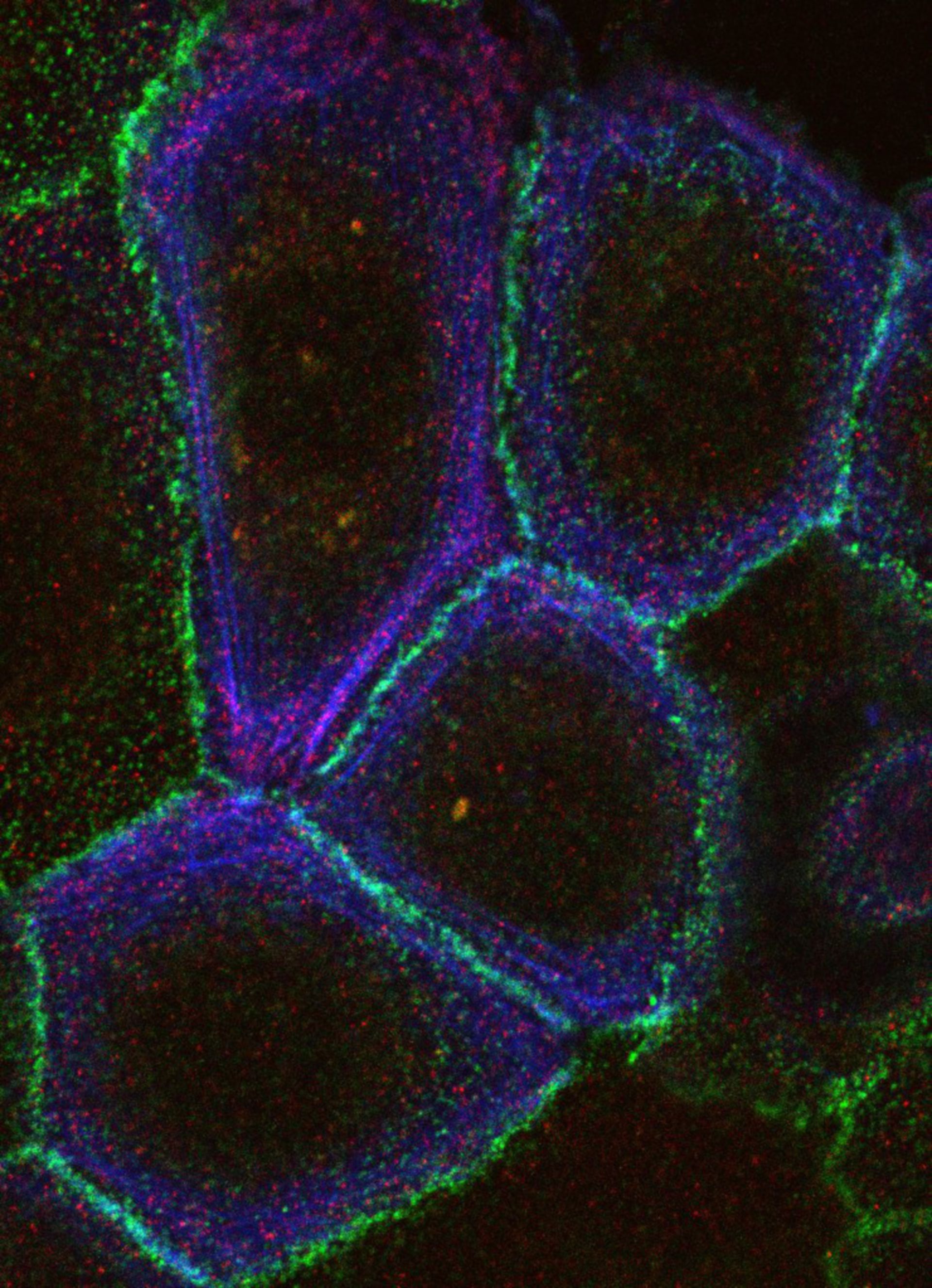
The relationship between the actin cytoskeleton (blue), adherens junctions (green) and phospho-Myosin Light Chain (MLC) (red) after 30 minutes of calcium stimulation.
Biological Process: Phosphorylation of myosin light chain, Calcium stimulation
The specimen was fixed with labelled with 4% paraformaldehyde and labeled with AlexaFluor488 phalloidin, rabbit anti alpha-catenin primary antibodies and Cy5 anti-rabbit secondary antibodies, mouse anti Phospho-MLC Ser18/Thr19 antibodies (Cell Signaling technologies) and mouse anti cy2 secondary antibodies. Images were acquired on a Zeiss LSM510 inverted microscope using a 63x 1.4 Numeric aperture oil immersion objective.
Author: Ann Wheeler
Source: The Cell: An Image Library