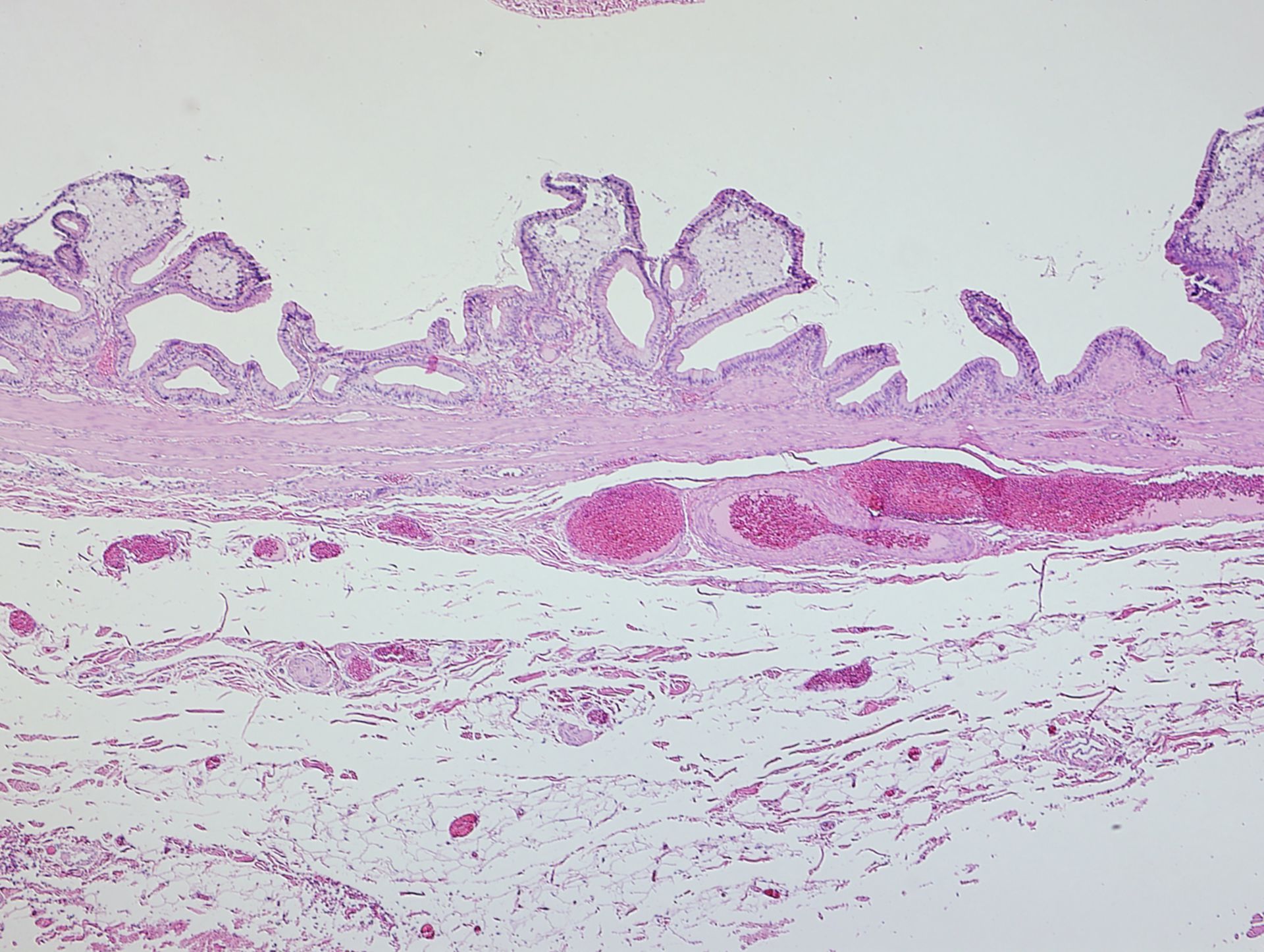
Gallbladder wall of a human patient with gallstones. This low magnification view shows the wall of the diseased gallbladder to have a top layer of highly polarized epithelial cells over a large volume of foamy macrophages laden with cholesterol. Below is muscle and mucous glands. The patient subsequently underwent gall bladder removal surgery.
Biological Process: Leukocyte chemotaxis involved in inflammatory response, Acute inflammatory response
Tissue was prepared by standard histological methods for pathology. Biopsy was fixed, cut, and stained with hematolylin and eosin. Images were collected using an Olympus BX51 scope, DP71 camera, and a 5x objective.
Author: William Karkow
Source: The Cell: An Image Library