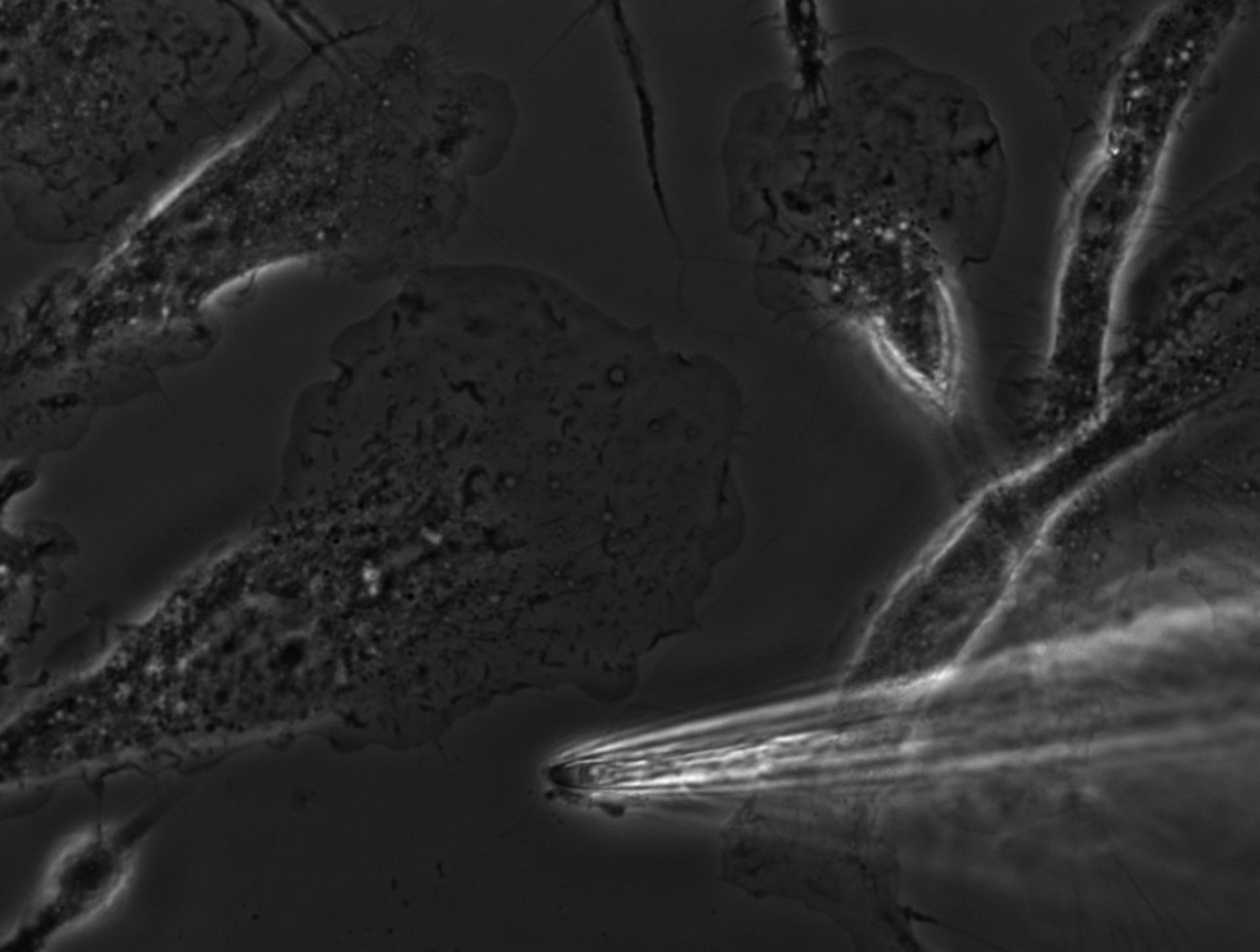
Primary bone marrow macrophages derived from mouse plated on a coverslip and imaged live. Phase contrast microscopy allows us to see the dense f-actin rich leading edge, ruffles, mitochondria, the nucleus, and other organelles throughout the volume of the cells. At the center of the image is a polarized lamellipod of a crawling cell. The glass micropipette at the bottom of the image is delivering CSF (colony stimulating factor) locally and the cell may be responding.
Biological Process: Ruffle organization
Live bone marrow macrophages plated in MatTek dish and imaged with a 60X N.A. 1.4 phase 3 Olympus objective on an IX71 microscope with a Cooke Sensicam QE camera and IPLab software running on a Dell Windows XP computer.
Author: Dianne Cox
Source: The Cell: An Image Library