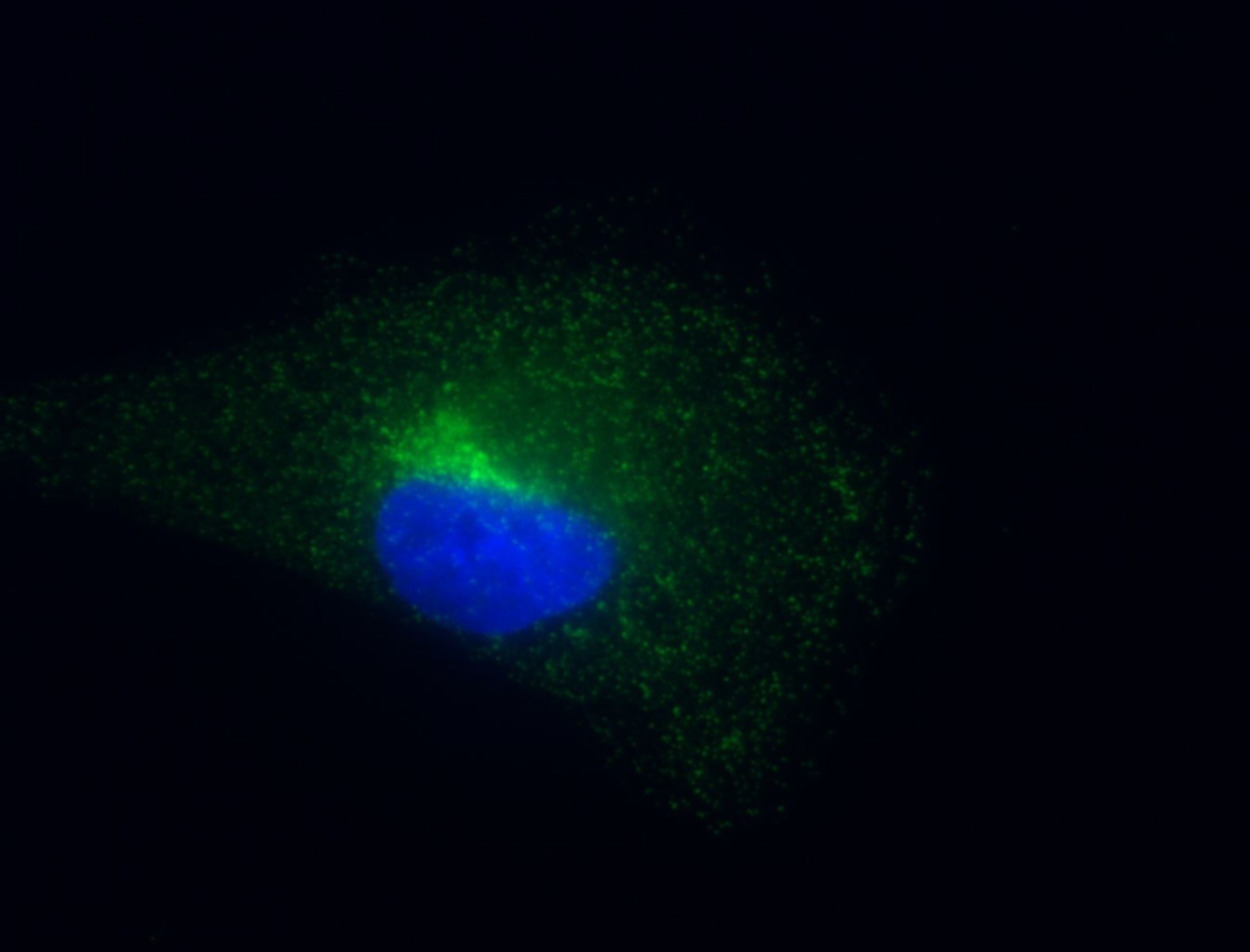
Cultured retinal pigment epithelial cells immunofluorescently labeled for clathrin (green) and nucleus (blue). The cells were fixed in 2% PFA and 0.5% Triton X-100 for 2 minutes followed by post-fixation 4% PFA. Clathrin was detected with X22 primary antibody and secondary FITC antibody. The nucleus was detected with DAPI staining. Images were collected on an Olympus IX-71 epifluorescence microscope using a 100X 1.4 NA objective with 4.500ms exposure for clathrin and 50ms exposure for DAPI (67nm/pixel).
Author: Sandra L. Schmid
Source: The Cell: An Image Library