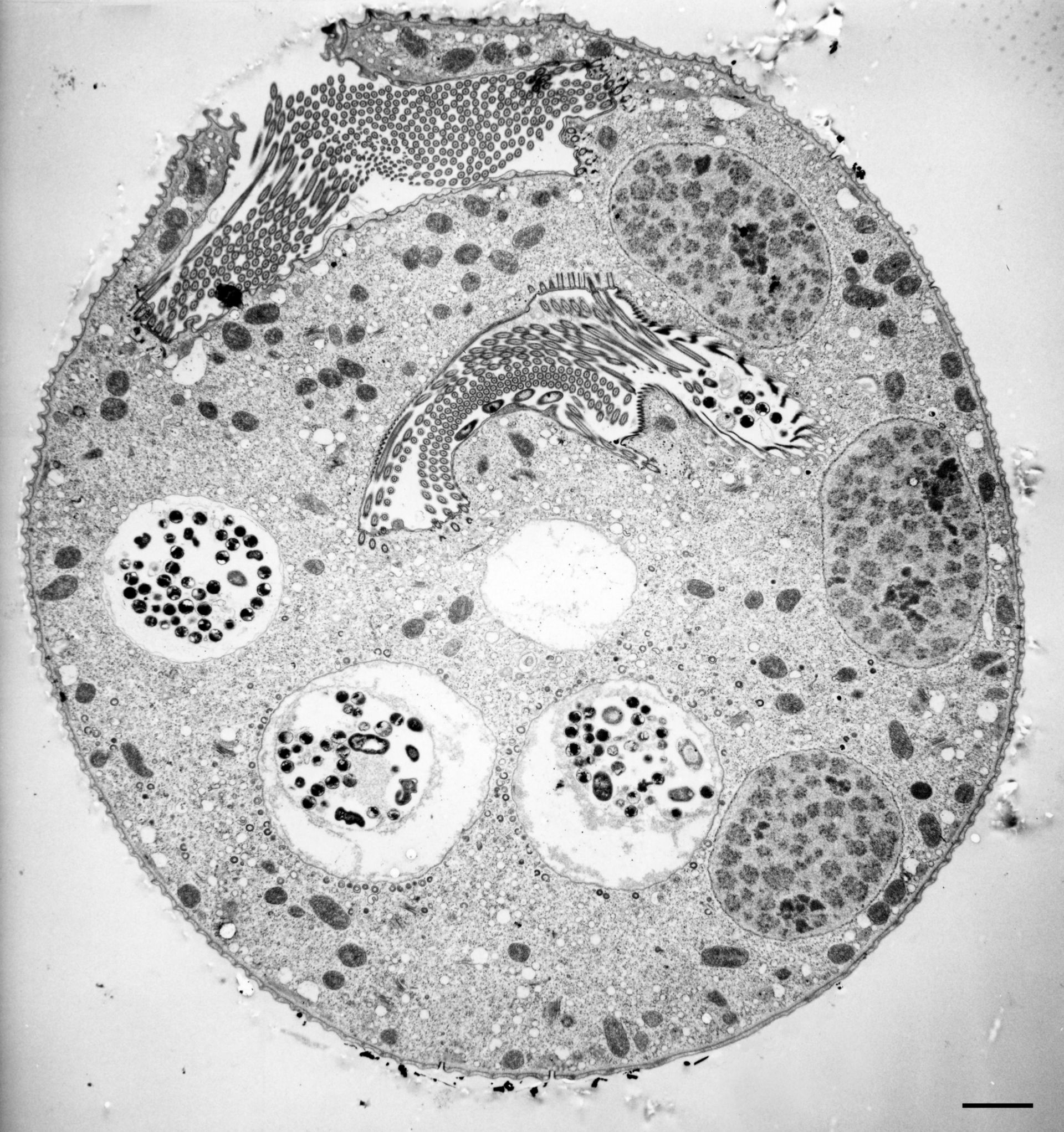
11 micrographs are views of a serially-sectioned contracted Vorticella convallaria cell that show the main features of this cell. This figure shows 4 food vacuoles; 3 segments of macronucleus; cytopharynx open to peristome; cytostome or cell mouth; perioral cilia covered by folds of the pellicle and cortex. TEM taken on 4/5/71 by R. Allen with Hitachi HU11A operating at 75kV. Neg. 2,150X. Bar = 2?m. The negative was printed to paper and the image was scanned to Photoshop. This digitized image is available for qualitative analysis. There is a high resolution version of this image in the library (CIL:39403) which is available for quantitative analysis. Additional information available at (http://www5.pbrc.hawaii.edu/allen/).
Biological Process: Digestive system process, Cytoplasm organization, Cortical cytoskeleton organization, Macronucleus organization, Oral apparatus organization, Digestive system process
Standard glutaraldehyde fixation followed by osmium tetroxide, dehydrated in alcohol and embedded in an epoxy resin. Microtome sections prepared at approximately 75nm thickness. Additional information available at (http://www5.pbrc.hawaii.edu/allen/).
Author: Richard Allen (University of Hawaii)
Source: The Cell: An Image Library