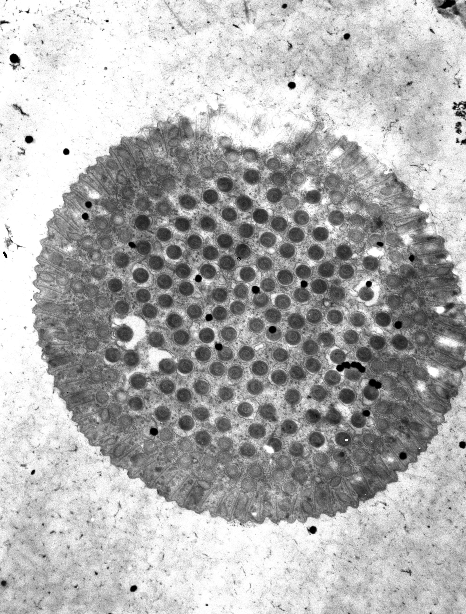
A transverse section near the tip of the oral proboscis of Didinium. This micrograph shows the extrusive organelles of the mucocysts adjacent to the plasma membrane, the toxicysts in a circular ring adjacent to the mucocysts, and the denser pexicysts in the central region of the proboscis. A section deeper in the proboscis is found at (CIL:17451). Further details available at Wessenberg, H. and Antipa, G. 1968. Studies on Didinium nasutum. I. Structure and ultrastructure. Protistologica 4:427-447. And details of the operation of this structure is available at Wessenberg, H. and Antipa, G. 1970. Capture and ingestion of Paramecium by Didinium nasutum. J.?Protozool. 17:250-270. TEM taken in 1966 by G. Antipa with JEM T6S operating at 50kV. Neg. 6,000X. The raw film was scanned with an Epson Perfection V750 Pro. This image is available for quantitative analysis. Standard glutaraldehyde fixation followed by osmium tetroxide, dehydrated in alcohol and embedded in an epoxy resin. Microtome sections prepared at approximately 50nm thickness.
Biological Process: Oral apparatus organization, Plasma membrane organization
Author: Gregory Antipa (San Francisco State University)
Source: The Cell: An Image Library