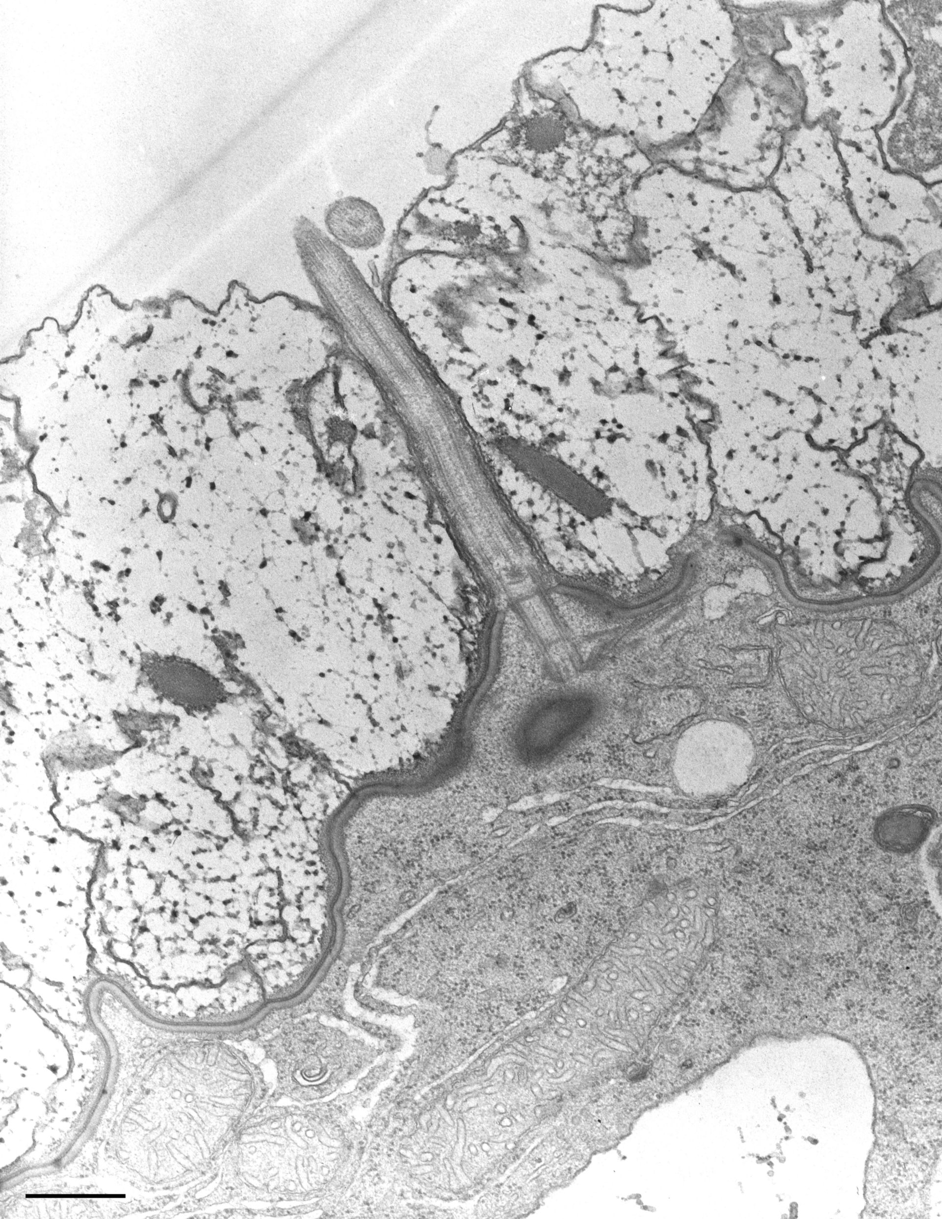
Detail of cell surface structures. The cell is covered by the plasma membrane and outer alveolar sac membrane which is separated from the inner alveolar sac membrane by an extensive alveolar space. Cilia extend through this alveolus to the true cell surface. TEM taken on 3/4/69 by R. Allen with Philips 300 operating at 60kV. Neg. 14,800X. Bar = 0.5?m. A print of the negative was scanned and processed in Photoshop. This image is best used for qualitative analysis. A high resolution image (CIL:7625) is available for quantitative analysis. Standard glutaraldehyde fixation followed by osmium tetroxide, dehydrated in alcohol and embedded in an epoxy resin. Microtome sections prepared at approximately 75nm. Additional information available at (http://www5.pbrc.hawaii.edu/allen/).
Biological Process: Cortical cytoskeleton organization, Plasma membrane organization, Ciliary or flagellar motility
Author: Richard Allen
Source: The Cell: An Image Library