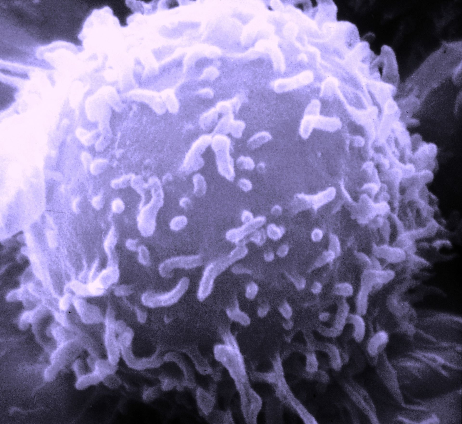
Lymphozyt (REM-Aufnahme, koloriert)
Lymphozyt unter dem Rasterelektronenmikroskop(Bildquelle:
Dr. Triche
National Cancer Institute)